Human Lipedema Adipocytes Show Metabolic Inflexibility Despite Preserved Adipogenesis
Poojari A, Dev K, Aguilan J, Hossain MA, Nguyen C, Al Ghadband S, Sidoli S, Rabiee A.

Our laboratory uses lipedema as a powerful disease model to ask broader questions about adipocyte identity, metabolic flexibility, mitochondrial adaptation, thermogenic signaling, and tissue communication.
Lipedema is a chronic adipose tissue disorder characterized by disproportionate, painful expansion of subcutaneous fat that is resistant to conventional weight-loss approaches. Our lab studies patient-derived adipose stem cells and differentiated adipocytes to determine why lipedema fat remains biologically and metabolically distinct from both lean and obesity-associated adipose tissue.
We focus on adipogenesis, lipid storage and mobilization, mitochondrial function, thermogenic responsiveness, secreted signaling, and the cellular programs that may contribute to the striking resistance of lipedema adipose tissue to metabolic remodeling.
Core question: what makes a lipedema adipocyte biologically distinct — and can that state be therapeutically reprogrammed?
Poojari A, Dev K, Aguilan J, Hossain MA, Nguyen C, Al Ghadband S, Sidoli S, Rabiee A.
Rabiee A. Frontiers in Cell and Developmental Biology.
View paper ↗Poojari A, Dev K, Rabiee A. Biomedicines.
View paper ↗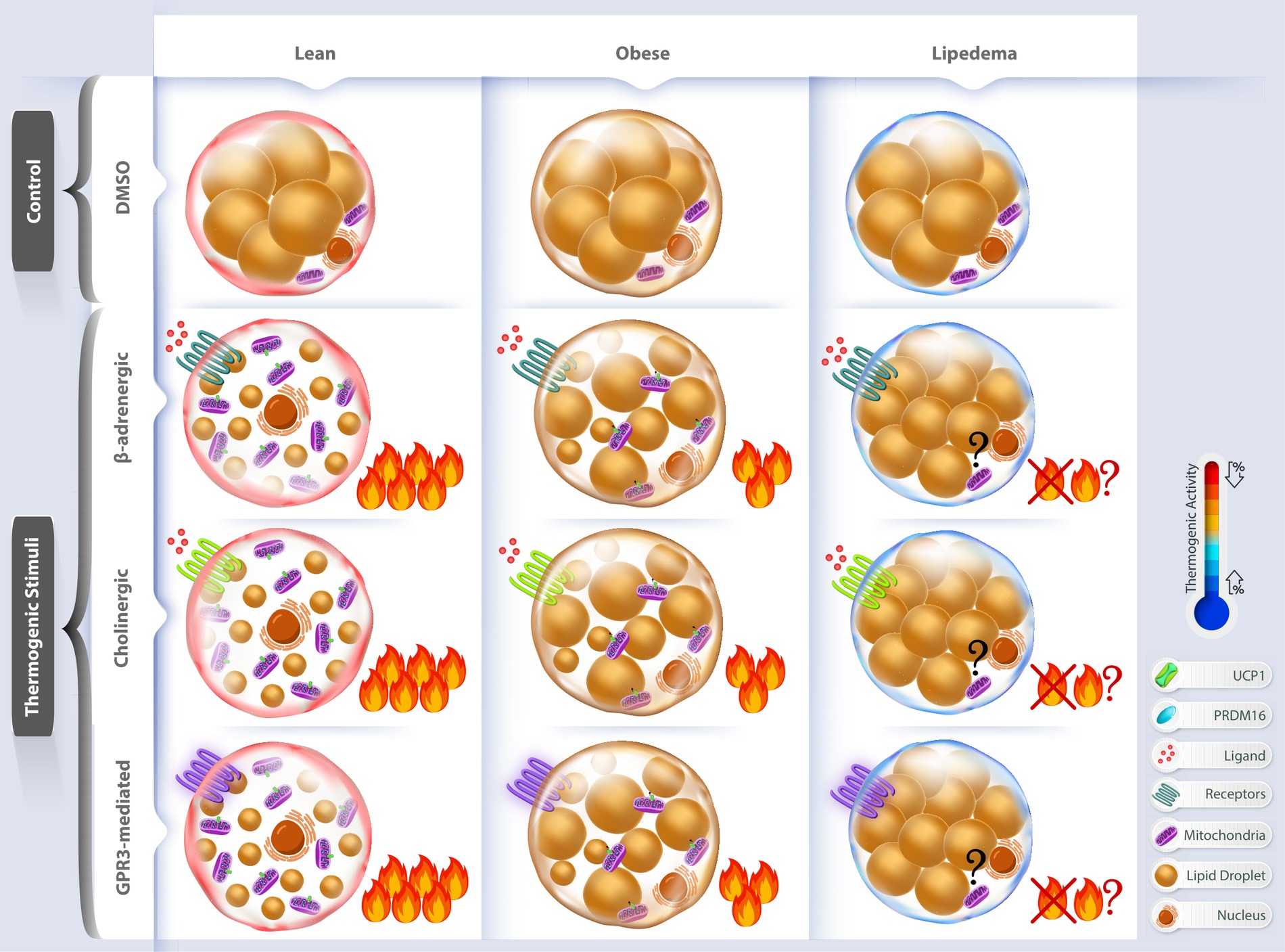
Mitochondria sit at the center of adipocyte metabolic flexibility. They determine whether fat cells can respond appropriately to changes in nutrient supply, energetic demand, thermogenic stimulation, and cellular stress.
Our lab studies how mitochondrial abundance, respiratory output, membrane potential, calcium handling, and structural remodeling become uncoupled in disease. We are particularly interested in the relationship between mitochondrial quantity and mitochondrial performance: an adipocyte may contain abundant mitochondrial machinery yet still fail to generate an appropriate energetic response.
We examine regulators of mitochondrial fusion and fission, oxygen-consumption responses, and signaling pathways that may explain why lipedema adipocytes retain biochemical capacity but remain functionally inflexible.
Core question: how does failure of mitochondrial adaptation lock adipocytes into a metabolically inflexible state?
Rabiee A, et al. Molecular Metabolism.
View paper ↗Rabiee A. Frontiers in Endocrinology.
View paper ↗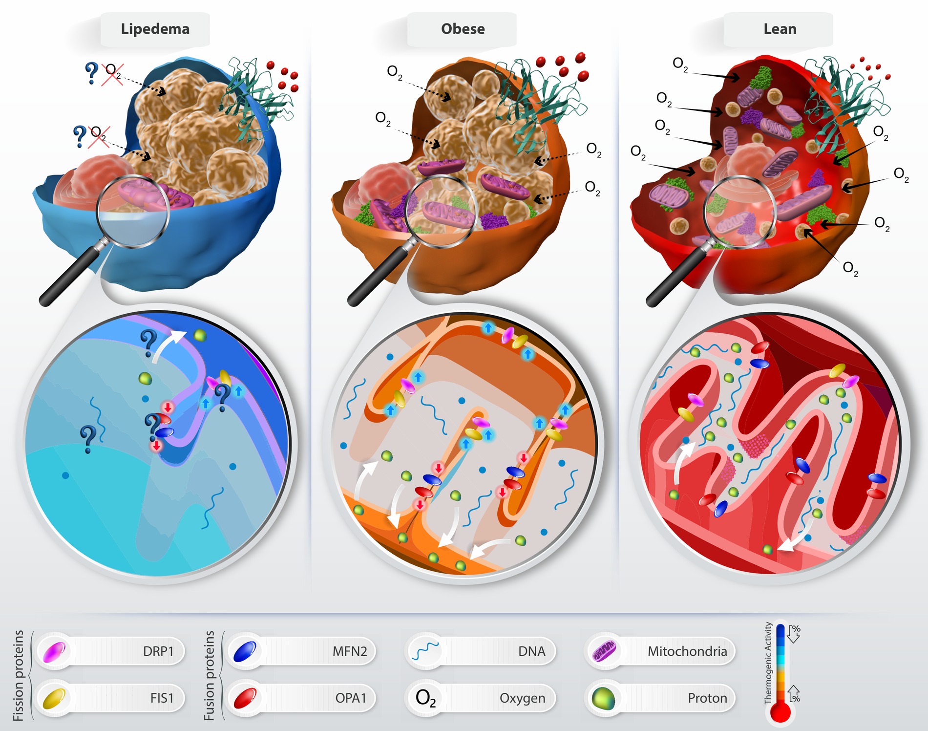
Adipocytes are not fixed in a single metabolic state. Under the right signals, they can alter lipid handling, mitochondrial activity, and thermogenic capacity. We study whether disease-associated adipocytes — particularly lipedema adipocytes — can be shifted toward a more metabolically active phenotype.
Our work compares classical adrenergic stimulation with alternative pharmacological strategies that may bypass impaired signaling pathways. Candidate compounds are evaluated for effects on lipid-droplet accumulation, lipolysis, thermogenic gene and protein expression, mitochondrial remodeling, membrane potential, and oxygen consumption.
Core question: can pharmacological reprogramming restore thermogenic responsiveness and metabolic flexibility in dysfunctional adipocytes?
Hossain MA, Poojari A, Rabiee A. Cells.
View paper ↗Brandao BB, Poojari A, Rabiee A. International Journal of Molecular Sciences.
View paper ↗Rabiee A. Frontiers in Endocrinology.
View paper ↗A newer direction in the laboratory extends our adipocyte biology program into perivascular adipose tissue (PVAT), where adipose dysfunction may directly influence vascular health.
We are developing studies focused on how PVAT changes across prediabetes and diabetes, with particular interest in sex-specific differences, adipose–vascular signaling, endoplasmic-reticulum stress, and calcium homeostasis.
This work is intentionally positioned as an emerging area that builds on our core expertise in adipocyte function, metabolic stress, and cellular signaling.
Emerging question: how does metabolic dysfunction in PVAT alter vascular function as diabetes develops?
Rabiee A, Rahimian R. Cells.
We are extending our adipocyte expertise to questions of tumor–stroma communication, including lipid transfer, metabolic adaptation, secreted factors, and the ways adipocyte phenotype may influence the tumor microenvironment.
This developing direction builds directly on the laboratory's strengths in adipocyte metabolism, lipid handling, live-cell analysis, and functional metabolic phenotyping.
Emerging question: how do adipocytes reshape the metabolic environment experienced by neighboring tumor cells?
We combine discovery-scale profiling with targeted functional assays to move from molecular signatures to testable mechanisms in adipocytes.
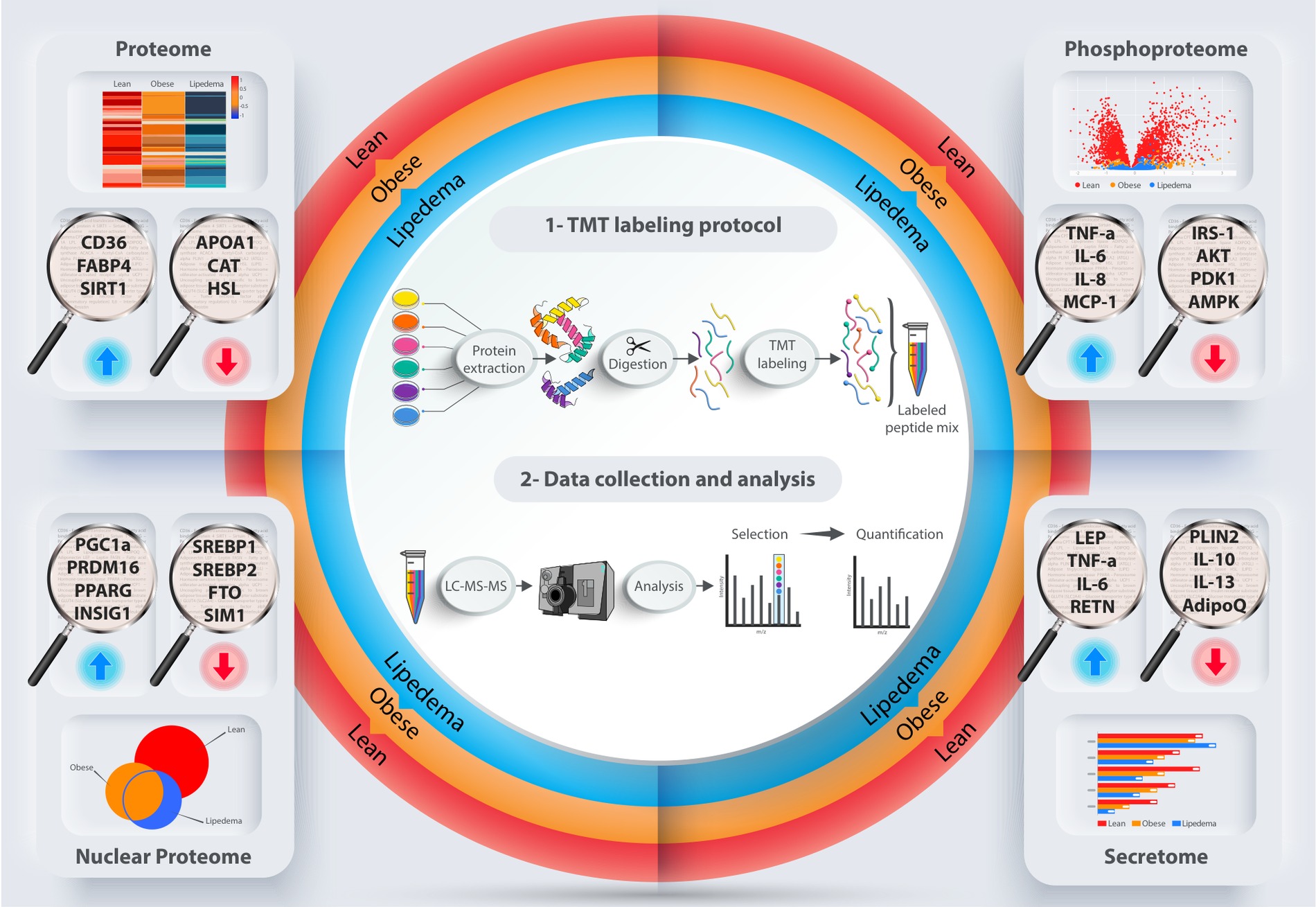
TMT proteomics, phosphoproteomics, nuclear proteomics, and secreted-protein analysis.
Oxygen-consumption profiling and functional assessment of mitochondrial adaptation.
CellCyte X imaging, lipid-droplet analysis, morphology, and dynamic cellular responses.
qPCR, Western blotting, pathway-focused validation, and mechanistic follow-up.
Human adipose-derived stem cells and differentiated adipocytes from lean, obese, and lipedema donors.
Mechanism-driven compound testing to probe thermogenic signaling and metabolic rescue.